RAM01 ANATOMICAL MODEL BRAIN
>Dissectible into halves
>Mounted on a base
>Natural or synthetic cast
>Different sections identified
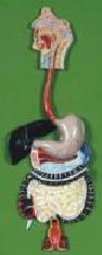
ANATOMICAL MODEL HUMAN DIGESTIVE SYSTEM
>Complete life-size unit
>Shows the alimentary canal up excretion
>Color feature finish to show inner organs
>Made from durable synthetic material
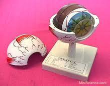
RAM03 ANATOMICAL MODEL EYE
>Enlarged 4 times
>Cross-section through the eye
>Shows sclera, choroid, retina optic nerve,
vitreous body, lens, iris, ciliary body and cornea
>Retina & choroidal vascular systems show
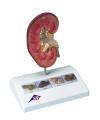
RAM05 ANATOMICAL MODEL KIDNEY
>Enlarged 4 times
>Dissected into halves
>Shows all the main features
>Made from durable synthetic material
>Mounted on a stand
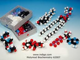
RAM010 MOLECULAR ATOMS MODEL
>Contain organic and inorganic sets
>Molecules range from simple to complex ions
>Gray links of 2 lengths for single and covalent
bonding
>Supplied with durable plastic tray
>Sphere:
#6 carbon, 22 oxygen, 10 nitrogen, 12 sulphur,
8 halogen, 14 metal, 7 P, 14 H
>Links:
#50 medium gray and mauve, 36 long gray
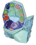
RAM08 ANATOMICAL ANIMAL CELL
The two piece animal cell model shows the form and structure of a typical animal cell as viewed by an electron microscope. For purposes of better illustration, all important organelles are shown in raised relief and displayed in color, e.g.:
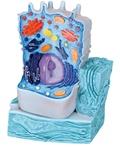
RAM08 ANATOMICAL PLANT CELL
>3 Dimensional >Shows form, structure, relationship of cell >Cross section mounted on plastic base >Fitted with transparent removable case >Numbered and color coded >Enlarged 20 000X >Shows centrioles, lysosomes, and fat vacuole
RAM ANATOMICAL MODEL: MITOSIS
The three-dimensional relief models are painted according to the usual coloring methods of microscopy, making the process of cell division easy to understand. The cell organelles are shown as if opened up in the lower part of the models. The models are equipped with magnets at the back so that for teaching purposes they can be easily arranged on a magnetic board in the classroom. The model series is supplied in a storage system (40 x 60cm) which can be fastened to the wall. A detailed description and handouts for your lessons are included.
1. Interphase
2. Prophase
3. Early prometaphase
4. Later prometaphase
5. Metaphase
6. Early anaphase
7. Later anaphase
8. Telophase
9. Cytokinesis
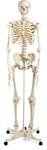
RAM06 ANATOMICAL MODEL SKELETON
>Full size model , fully artuculated
>Skull divided into 3 parts
>Arms and legs detachable
>Mounted on roller stand
>Height 1750mm
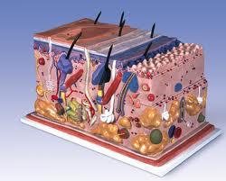
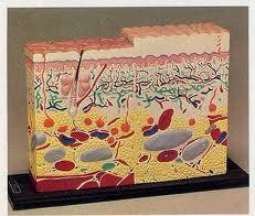
RAM011 ANATOMICAL SKIN MODEL
>Cut-away view shows skin layer, follicles,
sweat glands, nerve endings, blood vessels
>Each feature numbered, high impact plastic,
reinforced with foam.
> Enlarged cross section shows human skin
with 3 layers, front, side and back view,
mounted on plastic base,
>Not dissectible.
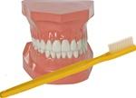
RAM ANATOMICAL DENTAL MODEL
>New and improved oversize >2X Study model and toothbrush set >Now completely unbreakable! >Fully functional
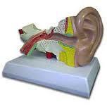
RAM02 ANATOMICAL MODEL EAR
>Four part model
>Made from rigid plastic
>Model enlarged 3 times
>Shows essential details of the ear
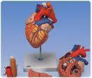
RAM04 ANATOMICAL MODEL HEART
>Enlarged 2.5 times
>Made from rigid plastic
>Dissectible into 3 parts
>Colour feature finish to show all parts
>Mounted on a stand
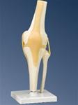
ANATOMICAL KNEE JOINT
This functional model provides a graphic demonstration of the anatomy and mechanics of the joint.
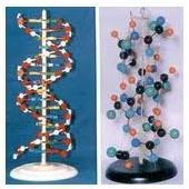
RAM09 ANATOMICAL DNA MODEL
>Simplified presentation of
double stranded DNA
> Durable plastic
> Colour coded to easily identify
>Height 500mm
>Contains 10 paired nucleotides
>Mounted on plastic base
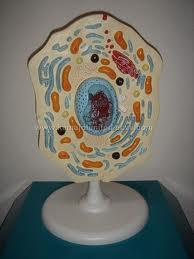
RAM08 ANATOMICAL HUMAN CELL
>3 Dimensional
>Shows form, structure, relationship of cell
>Cross section mounted on plastic base
>Fitted with transparent removable case
>Numbered and color coded
>Enlarged 20 000X
>Shows centrioles, lysosomes, and fat vacuoles
RAM ANATOMICAL MODEL: MEIOSIS
This newly developed 3B Scientific® meiosis model series shows the 10 stages of meiosis on the basis of a typical mammal cell at an enlargement of approximately 10,000 times. The detailed stages of meiosis
1. Interphase (stage of G1-phase)
2. Prophase I (leptotene)
3. Prophase I (zygotene and pachytene)
4. Prophase I (diplotene)
5. Prophase I (diakinesis)
6. Metaphase I
7. Anaphase I
8. Telophase I, cytokinesis I, interkinesis, prophase II and metaphase II
9. Anaphase II
10. Telophase II and cytokinesis II
The three-dimensional relief models are painted according to the usual coloring methods of microscopy, making the process of meiotic cell division easy to understand. The cell organelles are portrayed open in the lower part of the models. The meiosis models are equipped with magnets at the rear so that they can be easily arranged on a magnetic board in the classroom. The model series is supplied in a storage system (40 x 60 cm) that can be hung on a wall. The meiosis model comes complete with a detailed description and handouts to copy for your lessons.
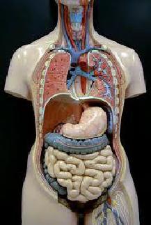
RAM07 ANATOMICAL MODEL TORSO
>Supplied as a complete piece
>Rubberised for durability
>Natural size
>Separable into 8 parts
>Mounted on stand
RAM12 ANATOMICAL SKIN MODEL
>Cut-away view shows skin layer, follicles,
sweat glands, nerve endings, blood vessels,
each feature numbered, high impact plastic,
reinforced with foam.
> Enlarged cross section shows human skin
with 3 layers, front, side and back view,
mounted on plastic base,
>Not dissectible.